– Professor and HLA Laboratory Director, University of Southern California and Children's Hospital Los Angeles, United States
Aim: Quantify HLA expression at the cellular level using imaging mass cytometry (IMC) to better understand the relationship between HLA expression and graft rejection.
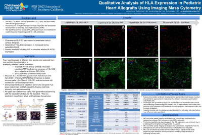
Methods: Four heart biopsies at different time points were selected from one pediatric heart recipient to exemplify different clinical scenarios: 1) states of both clinical antibody-mediated rejection (AMR) with strong presence of HLA-DQ donor-specific antibodies (DQ-DSA), 2) no AMR with presence of DQ-DSA. A 33-marker IMC panel was designed to detect immune cells, HLA-ABC, HLA-DR, and HLA-DQA. Regions of interest on paraffin-embedded biopsies were selected and combined on a single glass to reduce technical variability of IMC markers. Median molecular intensity (MMI) for each marker was normalized by min-max normalization with the 99% value set to 1 and the 1% percentile set to 0. Given that HLA class I is constitutively expressed across nearly all cells, we used HLA-DQ/HLA-ABC and HLA-DR/HLA-ABC ratios to evaluate relative levels of HLA expression in four heart biopsies. Variability in antibody binding and/or metal detection efficiency may influence signal strength. However, since these technical differences remain consistent across our four samples, relative expression was evaluated. Additionally, each biopsy contains many cell types, so observed differences may be influenced by variations in cellular composition. Ongoing single-cell analysis is underway to address this limitation.
Results: Across the four biopsies, both HLA-DR/HLA-ABC and HLA-DQ/HLA-ABC ratios varied, suggesting dynamic regulation of HLA antigen expression on heart allografts. Biopsy 1 served as a baseline with HLA-DR/HLA-ABC and HLA-DQ/HLA-ABC ratios at 1.07 and 0.53, respectively. Levels of HLA expression increased in biopsy 2 which was obtained during AMR (DR/ABC: 1.18; DQA/ABC: 0.79). HLA expression remained elevated in biopsies 3 and 4, with DR/ABC and DQ/ABC ratios of 1.25 and 0.64 in biopsy 3, and 1.46 and 0.89 in biopsy 4, respectively. Notably, biopsy 3 was obtained in the absence of AMR, while biopsy 4 was obtained during AMR.
Conclusion: These early results suggest that IMC and other spatial imaging technology may provide new insights into the relationship between HLA expression and AMR in allografts.
Footnotes: A subset of this data was presented at this years ISHLT meeting, but we expect to have updates by ASHI's meeting this October.

 (she/her/hers) photo")