– Assistant Director, University Health Network, Canada
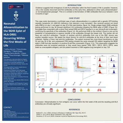
Body: Evidence suggests that emergence of anti-HLA antibodies within the first 6-weeks of life is possible. However, only a few studies document anti-HLA antibodies at this early time, attributed to either early allosensitization1 or via transplacental passage2. This is in contrast to the formation of ABO antibodies that occur between 3 and 6 months of age3. This case study documents a confirmed case of early allosensitization in a patient with a genetic ATP-binding cassette transporter A3 (ABCA3) deficiency that required a lung transplant. She received packed red blood cells (RBCs) on day 2, and again on day 25 of life (see timeline, figure 1a). Single antigen bead (SAB) antibody testing performed on day 36 showed significant HLA antibodies targeting the 96HK eplet of DRB1 (Figure 1b). Surrogate flow crossmatch with a donor that carried DR8 and DR14 resulted in a very strong B-cell crossmatch, confirming the specificity of the antibodies (Figure 1d). We performed SAB on the mother’s blood to rule out the possibility of transplacental or passive transfer of the antibodies through the breast milk. The mother did not have any anti-HLA antibodies. Transfer of residual anti-HLA antibodies from one of the RBC donors was another possible source. We tested the blood donors for anti-HLA antibodies at the time of their next blood collection. Neither of them had anti-HLA antibodies. The patient received two more blood transfusions on day 52 and day 58 of life, and subsequent PRA testing showed the same strength DR pattern targeting the 96HK eplet in SAB that also resulted in a positive B-cell flow crossmatch (Figure 1c,d). This observation suggests the antibodies were not acquired passively or they would have waned. DR8, DR11, DR12, DR13, DR14, were listed as unacceptable antigens, and the patient received a VXM negative lung transplant on day 120.
Conclusion: Allosensitization to HLA antigens can occur within the first week of life and the resulting anti-HLA antibodies are clinically significant.
Footnotes:
References: Bedford R., et al. (1993). Archives of Disease in Childhood, 68(1 Spec No), 49–51. Girelli, G., et al. (2015). Blood Transfusion 13(3), 484–497. Savulescu, D. M. et al. (2020). American Journal of Reproductive Immunology 84(2).